In this series of educational programs, MEDIVISION™ explores various imaging techniques and delivers in depth explanations of the technical process, equipment operations and general image interpretation as applied to each scanning technology.
A 7 part series introducing conventional, X-Ray and nuclear imaging in medical diagnostics.
This educational video series is still under production, please contact us or check back soon for more information on pricing and availability.
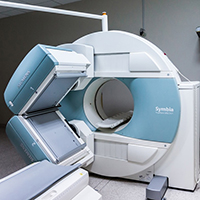
The first released title in our series, this program includes a brief history of the development of nuclear magnetic resonance and a Technical Aspects segment with an explanation of the interaction between magnetic fields and radiofrequency waves which produce MR images. The Operations segment looks at pre-MRI procedure screening and a current overview of protocol considerations as well as the positioning of various RF coils. Finally, in the Interpretation segment of the program we discuss the actual imaging sequences and provide general explanations of intensity for each sequence.

This program includes a brief history of the medical use of Ultrasound and an explanation of sound wave transmission, reception and image formation. Covers multiple ultrasound modulation techniques including Doppler, and the use of contrast agents. The Operations segment explains possible adverse effects along with an overview of interventional ultrasound, equipment adjustments and the use of ultrasound in obstetrics. The Interpretation segment of the program covers both B-scan and Doppler ultrasound.
X-Ray Imaging Breakout Series
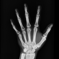
This program is the foundation of our 3-part breakout series on X-ray imaging. The history of X-radiation is covered from the invention of Geissler tubes and the work of Wilhelm Röntgen to the recognition of the dangers of exposure. We discuss the technical aspects of the modern x-ray machine and the imaging process, along with the introduction of digital radiography. The operations segment includes an overview of safety protocols, contrast agents and basic machine controls. Finally, guidelines for X-ray image interpretation are presented with basic radiographic opacities for various tissues and an explanation of Roentgen signs.
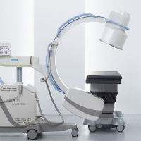
The second in our 3-part breakout series on X-ray imaging, Fluoroscopy covers the further development of x-ray imaging from the early discovery of its fluorescing properties to modern digital imaging with flat panel displays. The use of X-ray image intensifiers is explained, as well as an overview of their technical properties. Types of flat-panel detectors are discussed along with an overview of their mechanisms. The operations section includes further discussion of safety protocols, system configurations and general equipment adjustments, while the interpretations segment covers contrast agents and a brief discussion of image evaluation.
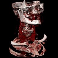
Beginning with a brief history of CT scanner development through the 1970's and 1980's, this program covers the technical aspects of x-radiation, image processing and construction as well as the Hounsfield unit scale. We discuss safety protocols including use of CT in pregnancy and in patients with implanted devices. Contrast agents are explained, along with a section on general usage protocols . The Interpretation segment of the program covers windowing along with artifacts and the means of minimizing them. The program concludes with a brief overview of new technology in computed tomography scanning.
Nuclear Medicine Breakout Series
This program, the first in a two part segment on nuclear medicine, includes a brief history of Positron-Emission Tomography in medical imaging and an explanation of radiotracers and the means of their detection in the imaging process. The Operations segment discusses preparation of the radiotracers along with overviews of attenuation correction, 2D and 3D reconstruction and "Time of Flight" technique. We describe the applications of PET in Oncology, Neuroimaging, Cognitive Neuroscience and other areas of diagnosis and research.
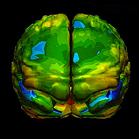
The second program in our nuclear medicine segment, this presentation describes the defining moments in the history of SPECT and describes the gamma camera imaging process. The advantages of integrated SPECT/CT scanning are discussed, along with the more commonly used radiotracers. The Operations segment discusses general protocols and provides overviews of the current applications of SPECT in myocardial perfusion imaging, functional brain imaging, diagnosis of infection and inflammation and oncology. Finally, the interpretation segment of the program covers examples of SPECT/CT imaging in various cerebrovascular diseases and dementias as well as the myocardial perfusion study.